I. Introduction
Magnetic Resonance Imaging (MRI) technology has transformed the medical landscape, offering an unprecedented glimpse into the human body. This non-invasive diagnostic tool uses magnetic fields and radio waves to produce high-resolution images of internal structures, revolutionizing the way healthcare professionals diagnose and treat various medical conditions. Medical imaging plays a vital role in modern healthcare, enabling early detection, accurate diagnosis, and effective treatment of diseases. MRI, in particular, has emerged as a game-changer, providing unparalleled insights into the body’s intricate workings. This essay explores the impact of MRI on medical diagnostics, highlighting its history, principles, applications, advantages, and recent advancements, demonstrating how MRI has revolutionized our understanding of the human body.
II. History of MRI
The development of Magnetic Resonance Imaging (MRI) technology is a story of innovation and collaboration among scientists and researchers across various disciplines. The roots of MRI can be traced back to the 1940s, when physicists Felix Bloch and Edward Purcell discovered the phenomenon of nuclear magnetic resonance (NMR). This discovery led to the development of NMR spectroscopy, a technique used to study the properties of atomic nuclei.
In the 1950s and 1960s, scientists like Richard Ernst and Raymond Damadian expanded on this research, exploring the potential of NMR for medical applications. Damadian, in particular, is credited with inventing the first MRI machine in 1971, which used a strong magnetic field to align hydrogen nuclei in the body and produce a signal.
The 1970s and 1980s saw significant advancements in MRI technology, with the introduction of new techniques like spin density and magnetic field gradients. This led to the development of the first commercial MRI scanners, which were large, cumbersome, and expensive. Despite these limitations, MRI quickly gained recognition for its ability to produce high-resolution images of internal structures without the use of ionizing radiation.
In the 1990s and 2000s, MRI technology underwent rapid evolution, with the introduction of new sequences like functional MRI (fMRI), magnetic resonance angiography (MRA), and magnetic resonance spectroscopy (MRS). These advancements enabled researchers to study brain function, blood flow, and metabolic processes in unprecedented detail.
Today, MRI machines are an essential tool in hospitals and research institutions worldwide, with ongoing innovations like high-field MRI, ultra-high field MRI, and artificial intelligence-powered image analysis pushing the boundaries of medical diagnostics and research.
The history of MRI is a testament to human ingenuity and the power of interdisciplinary collaboration, demonstrating how fundamental scientific discoveries can lead to life-changing medical breakthroughs.
III. How MRI Works
Have you ever wondered how MRI machines produce those incredibly detailed images of our internal organs and structures? It’s actually pretty fascinating!
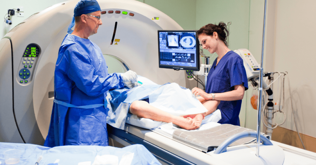
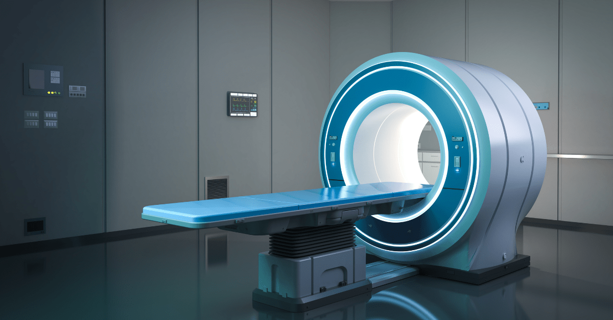
The process begins when you lie down inside the MRI machine, which is essentially a giant magnet. This magnet creates a strong magnetic field that aligns the hydrogen atoms in your body. Yes, you read that right – hydrogen atoms! They’re the most abundant atoms in our bodies, and they’re capable of being magnetized.
Once the hydrogen atoms are aligned, a radio wave pulse is sent through your body. This pulse disturbs the alignment of the atoms, causing them to emit signals, which are then picked up by the MRI machine. These signals are used to create detailed images of your internal structures.
But here’s the really cool part: the MRI machine can differentiate between different types of tissue based on their magnetic properties. For example, it can tell the difference between fat, water, and other tissues. This is because each type of tissue responds differently to the magnetic field and radio waves.
The MRI machine uses a combination of three main components to produce images:
- Main Magnet: This is the strong magnetic field that aligns the hydrogen atoms.
- Gradient Coils: These coils create spatial gradients in the magnetic field, allowing the machine to pinpoint the exact location of the signals.
- Radio Frequency Coils: These coils send and receive the radio wave pulses that disturb the alignment of the atoms and detect the signals.

The signals are then processed using a technique called Fourier transform, which converts the raw data into detailed images. These images can be viewed in multiple planes and dimensions, allowing radiologists and doctors to examine your internal structures from all angles.
It’s truly mind-boggling to think about how MRI machines can produce such detailed images of our bodies without even touching us! The technology has come a long way since its inception, and it continues to evolve, enabling us to better understand the human body and diagnose medical conditions more effectively.
IV. Applications of MRI
As a doctor, I can attest that MRI has revolutionized the way we diagnose and treat various medical conditions. The technology has been a game-changer in many fields, including neurology, cardiology, oncology, and orthopedics. Let me share some examples of how MRI has impacted our practice:
Neurology
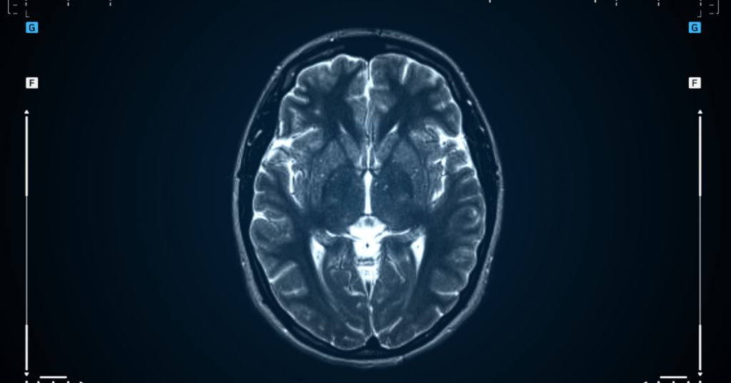
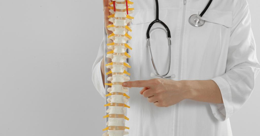
MRI has enabled us to visualize the brain and spinal cord in unprecedented detail. We can diagnose conditions like multiple sclerosis, Parkinson’s disease, and brain tumors with greater accuracy. Functional MRI (fMRI) even allows us to map brain activity, helping us understand brain function and behavior.

Cardiology
MRI has improved our understanding of heart structure and function. We can diagnose conditions like coronary artery disease, cardiomyopathy, and congenital heart defects with greater precision. MRI also helps us monitor the effectiveness of treatments like angioplasty and heart transplantation.
Oncology
MRI plays a crucial role in cancer diagnosis and treatment. We can detect tumors and metastases earlier and more accurately than ever before. MRI-guided biopsies enable us to target specific areas for tissue sampling, reducing the risk of false negatives.
Orthopedics
MRI has transformed the way we diagnose and treat musculoskeletal conditions. We can visualize joint and muscle structures in high detail, allowing us to diagnose conditions like ligament tears, tendonitis, and osteoarthritis more effectively.
Other Applications
MRI is also used in:
- Pediatrics: to diagnose conditions like cerebral palsy and hydrocephalus
- Gastroenterology: to evaluate liver and pancreatic function
- Urology: to diagnose prostate cancer and kidney stones
- Research: to study brain development, aging, and various diseases
In conclusion, MRI has become an indispensable tool in modern medicine. Its versatility, sensitivity, and specificity have improved patient outcomes and saved countless lives. As a doctor, I’m grateful for the insights MRI provides, enabling me to provide better care for my patients.
V. Advantages and Limitations
As with any medical imaging modality, MRI has its advantages and limitations. Understanding these factors is crucial for effective patient care and research applications.
Advantages:
- High-resolution imaging: MRI provides exceptional spatial resolution, allowing for detailed images of internal structures.
- Non-invasive: MRI is a non-invasive procedure, eliminating the risk of complications associated with invasive procedures.
- No radiation exposure: MRI does not use ionizing radiation, making it a safer option for patients, especially children and pregnant women.
- Multi-planar imaging: MRI allows for imaging in multiple planes, enabling a more comprehensive understanding of complex anatomical structures.
- Functional imaging: MRI can assess organ function, blood flow, and metabolic activity, providing valuable information for diagnosis and treatment.
- High sensitivity and specificity: MRI has high sensitivity and specificity for detecting various conditions, reducing false positives and false negatives.
Limitations:
- Claustrophobia: Some patients may experience claustrophobia or anxiety during the MRI procedure, which can be managed with sedation or open-bore machines.
- Cost and availability: MRI machines are expensive, and access may be limited in resource-constrained settings.
- Image artifacts: MRI images can be affected by motion artifacts, magnetic field inhomogeneities, and other technical factors.
- Contrast agent risks: Gadolinium-based contrast agents used in MRI can cause adverse reactions, such as nephrogenic systemic fibrosis.
- Pregnancy and breastfeeding: While MRI is generally safe during pregnancy and breastfeeding, caution is advised, and consultation with a radiologist is recommended.
- Metallic implant compatibility: MRI is contraindicated in patients with certain metallic implants, such as pacemakers, cochlear implants, and ferromagnetic materials.
By understanding the advantages and limitations of MRI, healthcare professionals can optimize its use, minimize risks, and provide better patient care.
VI. Recent Advancements and Innovations
Magnetic Resonance Imaging (MRI) technology has undergone significant advancements in recent years, leading to improved image quality, faster acquisition times, and new applications. Some of the recent developments and innovations include:
- High-Field MRI: The introduction of high-field MRI machines (7.0 Tesla and above) has enabled higher spatial resolution, improved signal-to-noise ratios, and better diagnostic accuracy.
- Functional MRI (fMRI): Advancements in fMRI have enabled researchers to study brain function, neural connectivity, and cognitive processes in greater detail.
- Diffusion Tensor Imaging (DTI): DTI has improved the visualization of white matter tracts in the brain, enabling better diagnosis and treatment of neurological conditions.
- Magnetic Resonance Spectroscopy (MRS): MRS has been enhanced to provide more accurate information on metabolic processes, allowing for earlier detection and monitoring of diseases.
- Parallel Imaging: Parallel imaging techniques have significantly reduced acquisition times, making MRI more accessible and patient friendly.
- Artificial Intelligence (AI) and Machine Learning (ML): AI and ML algorithms are being integrated into MRI to improve image reconstruction, automate image analysis, and aid in diagnosis.
- Open-Bore and Wide-Bore MRI: Open-bore and wide-bore MRI machines have been designed to reduce claustrophobia and accommodate larger patients.
- Portable and Low-Field MRI: Portable and low-field MRI machines are being developed for point-of-care imaging, emergency situations, and resource-constrained settings.
- MRI-Guided Interventions: MRI is being used to guide minimally invasive procedures, such as biopsies and tumor treatments, with greater precision and accuracy.
- Research and Development: Ongoing research is focused on developing new MRI sequences, improving image resolution, and expanding applications in fields like cardiology, oncology, and pediatrics.
These advancements and innovations have transformed the field of MRI, enabling healthcare professionals to provide better patient care, improve diagnostic accuracy, and drive medical research forward.
VII. Conclusion
Magnetic Resonance Imaging (MRI) has revolutionized the field of medical imaging, offering unparalleled insights into the human body. From its inception to the present day, MRI technology has undergone significant advancements, transforming the way we diagnose and treat various medical conditions.
In this essay, we have explored the history of MRI, its principles, applications, advantages, limitations, and recent innovations. We have seen how MRI has impacted various fields, including neurology, cardiology, oncology, and orthopedics, and how it has improved patient outcomes and saved countless lives.
As we move forward, it is essential to continue investing in MRI research and development, addressing the limitations and challenges associated with this technology. The integration of artificial intelligence, machine learning, and other cutting-edge technologies will further enhance the capabilities of MRI, enabling us to tackle complex medical challenges and improve human health.
In conclusion, MRI is a testament to human ingenuity and the power of medical innovation. Its impact on modern medicine is undeniable, and its potential for future breakthroughs is vast. As we continue to push the boundaries of MRI technology, we can expect even more remarkable advancements, leading to better patient care, improved diagnostic accuracy, and a brighter future for humanity.
Final Thoughts
- MRI has transformed medical imaging, offering high-resolution images and functional insights.
- Continued innovation and research are crucial for addressing limitations and unlocking new applications.
- Integration with AI, ML, and other technologies will further enhance MRI capabilities.
- MRI has the potential to tackle complex medical challenges and improve human health.
- Its impact on modern medicine is undeniable, and its future prospects are vast.
Further Reading
- Recent Advances in MRI Technology
- MR Imaging in the 21st Century: Technical Innovation over the First Two Decades
- The Future of MRI: Emerging Technologies and Applications
Explore Micro2media.com
- Green Choices Go High-Tech: How Eco-Friendly Products are Shaping the Future with ICT Innovation
- Unlocking the Skin-Boosting Benefits of Shea Butter: A Comprehensive Guide
- Sleepless in Seattle (and Everywhere Else): Conquering Chronic Insomnia

